Filter articles
标签
产品
Loading...
![[Translate to chinese:] Colon adenocarcinoma and normal colon at the tumor margin. 13 biomarkers shown including Cadherin, CD3, CD4, CD8, CD20, CD31, CD45, Collagen, Caspase 9, BCL2, Beta-Catenin, Vimentin, and Smooth Muscle Actin. [Translate to chinese:] Colon adenocarcinoma and normal colon at the tumor margin. 13 biomarkers shown including Cadherin, CD3, CD4, CD8, CD20, CD31, CD45, Collagen, Caspase 9, BCL2, Beta-Catenin, Vimentin, and Smooth Muscle Actin.](/fileadmin/_processed_/2/9/csm_Colon_Adenocarcinoma_and_Normal_Colon_13_Markers_1be18263b2.jpg)
Uncover the Hidden Complexity of Colon Cancer with Big Data
Colorectal cancer poses a significant health burden. While surgery is effective initially, some patients develop recurrent secondary disease with poor prognosis, necessitating advanced therapies like…
Loading...
![[Translate to chinese:] Digital microscopy simplifies documenting cell-culture results electronically while following 21 CFR part 11 guidelines for biopharma. [Translate to chinese:] Digital microscopy simplifies documenting cell-culture results electronically while following 21 CFR part 11 guidelines for biopharma.](/fileadmin/_processed_/9/6/csm_Documentation_of_cell-culture_results_with_Mateo_FL_following_21_CFR_part_11_guidelines_cfc8cdecb7.jpg)
Introduction to 21 CFR Part 11 for Electronic Records of Cell Culture
This article provides an introduction to the recommendations of 21 CFR Part 11 from the FDA, specifically focusing on the audit trail and user management in the context of cell-culture laboratories.…
Loading...
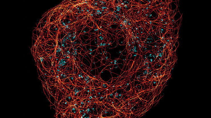
A Guide to Super-Resolution
Find out more about Leica super-resolution microscopy solutions and how they can empower you to visualize in fine detail subcellular structures and dynamics.
Loading...
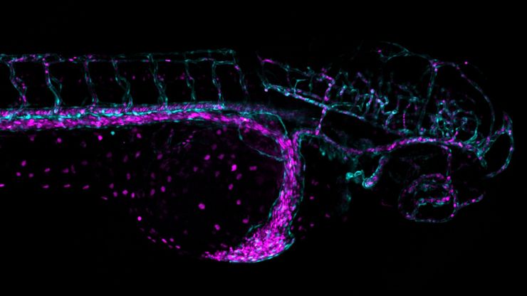
Overcoming Challenges with Microscopy when Imaging Moving Zebrafish Larvae
Zebrafish is a valuable model organism with many beneficial traits. However, imaging a full organism poses challenges as it is not stationary. Here, this case study shows how zebrafish larvae can be…

