Filter articles
标签
产品
Loading...
.")
双视野光片显微镜,适用于大型多细胞系统
展示复杂多细胞系统的动态是生物学中的一个基本目标。为了应对在大型时空尺度上进行活体成像的挑战,作者在《自然·方法》杂志上发表的一篇论文中介绍了一种开放式多样本双视野光片显微镜。研究发现,Viventis LS2 Live显微镜在以单细胞分辨率成像大型样本方面取得了显著进展。


Loading...
 and mito OM (red) in a live U2OS cell")
多色四维超分辨光片显微镜
人工智能显微术研讨会主要关注和讨论显微术和生物医学成像领域的最新人工智能技术和工具。在该科学演示中,Yuxuan Zhao展示了如何通过渐进式深度学习策略并结合“双环调制的SPIM”设计改善活细胞中的细胞器三维成像。
Loading...

利用DLS对细胞球中的抗癌药物摄取进行成像
细胞球3D细胞培养模型模拟了活组织的生理和功能,使其成为研究肿瘤形态和筛选抗癌药物的有用工具。药物AZD2014是一种公认的哺乳动物雷帕霉素靶蛋白(mTOR)通路抑制剂[1]。mTOR的异常激活会促进肿瘤生长和转移,导致AZD2014进入临床试验作为抗癌分子。其具体的抗肿瘤机制尚不清楚。
Loading...
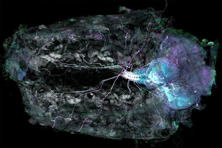
Understanding Motor Sequence Generation Across Spatiotemporal Scales
We have developed a microscopy-based pipeline to characterize a developmentally critical behavior at the pupal stage of development, called the ecdysis sequence. We study brain-wide neuronal activity…
Loading...
![3D glomeruli in a portion of an ECi-cleared kidney scanned by light sheet microscopy. Courtesy of Prof. Norbert Gretz, Medical Faculty Mannheim, University of Heidelberg [1].](/fileadmin/_processed_/d/d/csm_DLS-Sample-Preparation-Intr_915e0fd7c2.jpg "3D glomeruli in a portion of an ECi-cleared kidney scanned by light sheet microscopy. Courtesy of Prof. Norbert Gretz, Medical Faculty Mannheim, University of Heidelberg [1].")
使用安装框架进行光片样品准备
样品处理通常是光片显微镜研究中的一个关键话题。徕卡显微系统的TCS SP8 DLS将光片技术集成到倒置共聚焦平台中,因此可以利用关于样品安装和XY-stage功能的一般原则。本文将描述一组安装框架,这些框架不仅允许准备更多的样品,尤其是在使用诸如BABB(苯甲醇苯甲酸酯)等潜在有害的安装介质时,亦具有广泛的适用性。
Loading...

使用旋转设备进行光片样本安装
TCS SP8 DLS 显微镜采用了一种创新的设计理念,将光片显微技术集成到共聚焦显微镜中。得益于其独特的光学架构,样本可以像进行标准共聚焦成像一样,安装在标准玻璃底培养皿上。与传统的样本安装程序相比,这一过程只需进行少量调整。
Loading...

使用 U 形玻璃毛细管进行样品装载
徕卡显微系统的DLS显微镜系统是一种创新概念,将光片显微技术集成到共聚焦平台中。由于其独特的光学结构,样本可以安装在标准玻璃底培养皿上,与传统的安装程序相比,几乎不需要或只需很少的适应。在这里,我们介绍了一种便捷的方法,能够快速准备样本以进行光片成像。
