
Filter articles
标签
产品
Loading...

超分辨率显微镜图片库
由于光的衍射极限,传统共聚焦显微镜无法分辨约240纳米以下的结构。当需要提高分辨率以研究衍射极限尺度以下的结构和分子事件时,会使用超分辨率显微镜技术,如STED、PALM或STORM,或某些解卷积处理方法。
Loading...

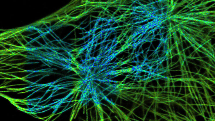
细胞活成像的纳米级扩展
新的STED显微技术方法——TauSTED Xtend,使得在纳米级别下对活体完整样本进行扩展多色成像成为可能。通过结合空间和寿命信息,TauSTED Xtend提供了额外一层信息,允许在极低的光剂量下分辨小细节并在整体结构中解析它们。
Loading...
, actin network (ATTO 647N), and nuclear pore basket (CF 680R).")
STED样品制备指南
这份指南旨在帮助用户优化受激发射损耗(STED)纳米成像的样品制备,特别是在使用徕卡微系统的STED显微镜时。它提供了单色STED成像用荧光标记的概述,并对其性能进行了评级。


Loading...
通过非拟合且简便的 FRET-FLIM 方法可视化蛋白质 - 蛋白质相互作用
了解活细胞中的分子相互作用对于解读大多数细胞功能背后的分子机制至关重要。研究蛋白质-蛋白质相互作用的金标准是福斯特共振能量转移(FRET)。尽管有几种方法可以在生物样品中证明FRET,但使用荧光寿命成像显微镜(FLIM)可以基于仅供体荧光的行为直接量化FRET。
Loading...

通过 11 种颜色的光谱分离实现超多标记
荧光显微镜是生命科学研究的基本工具,随着细胞组织和模式生物多色标记策略的发展而不断发展和成熟。分子特异性标记多种物种的能力需要适当的工具来识别样品中的多种荧光标签。严格分离多个标签对于获得有意义的成分、丰度、结构和功能读数至关重要。这种所谓的超多标技术在揭示组织组织、癌症进展、肿瘤免疫相互作用和传染病机制等关键方面已变得十分突出。
