
Filter articles
标签
产品

Loading...
Understanding Motor Sequence Generation Across Spatiotemporal Scales
We have developed a microscopy-based pipeline to characterize a developmentally critical behavior at the pupal stage of development, called the ecdysis sequence. We study brain-wide neuronal activity…
Loading...

使用增强功能电子显微镜研究大脑切片中的突触
神经科学的一个基本问题就是突触的结构与其功能特性之间有何关系?过去几十年,电生理学揭示了突触传递机制,而电子显微镜(EM)深入探索了突触形态。用于关联突触生理学和超微结构的方法可以追溯到20世纪中叶。目标是获得突触传递的快照,即捕获电子显微照片中的动态过程。
Loading...
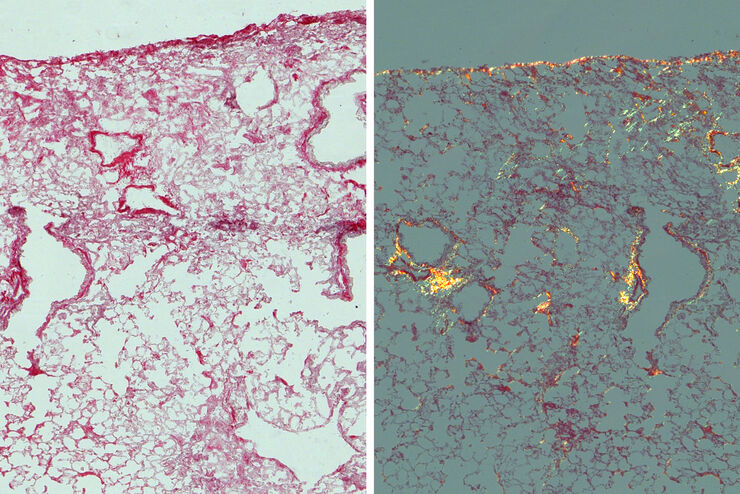
肺纤维化研究
本文中所示结果表明,相比明场,使用偏振光可以更清晰地分辨小鼠肺组织中的胶原蛋白纤维化和非纤维化区域。为更好地理解促使瘢痕组织形成的肺纤维化,正常情况下会研究组织中的纤维化区域。分别使用明场和偏振光对小鼠肺组织中的胶原蛋白纤维化病变区域进行成像。成像胶原蛋白时,使用一般的染色法和明场显微镜很难区分纤维化和非纤维化区域。本文中使用偏振光对肺组织进行成像,两个区域呈现出非常明显的颜色差异。
Loading...

Image Gallery: THUNDER Imager
To help you answer important scientific questions, THUNDER Imagers eliminate the out-of-focus blur that clouds the view of thick samples when using camera-based fluorescence microscopes. They achieve…
Loading...

An Introduction to Computational Clearing
Many software packages include background subtraction algorithms to enhance the contrast of features in the image by reducing background noise. The most common methods used to remove background noise…
Loading...
Computational Clearing - 增强3D标本成像
本次网络研讨会旨在阐明有助于THUNDER显微成像仪实现三维样品可视化的关键规格,并改进研究人员的成像相关工作流程。
Loading...
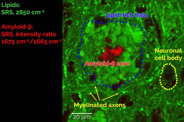
受激拉曼散射显微镜探测神经退行性疾病
Despite decades of research, the molecular mechanisms underlying some of the most severe neurodegenerative diseases, such as Alzheimer’s or Parkinson’s, remain poorly understood. The progression of…
Loading...

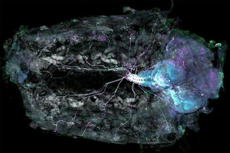
斑马鱼大脑高分辨率全器官成像
结构信息是理解复杂生物系统的关键,而脊椎动物的中枢神经系统是最复杂的生物结构之一。要想从发育中的斑马鱼身上分离出一个完整的大脑,我们需要覆盖大约10平方毫米的区域,深度在毫米范围内。通常,低倍透镜不能提供足够的分辨率来揭示神经组织中复杂结构之间的相互作用。此外,由于散射过程,使用共聚焦显微镜在致密生物组织内成像深度通常限制在大约10微米。
