
Filter articles
标签
 and oblique (right) brightfield illumination using a Leica compound microscope. The defect on the wafer surface is clearly more visible with oblique illuminati")
Loading...
 of an image of two points where the distance between them corresponds to the Rayleigh criterion.")
显微镜分辨率:概念、因素和计算
在显微镜学中,‘分辨率’一词用于阐述显微镜对细节进行区分的能力。换言之,这是样本内两个能被观察人员或者显微镜摄像头区分的实体点之间的最小距离。显微镜的分辨率本质上与光学元件的数值孔径(NA)以及用于观察样本标本的光波长有关。此外,我们必须考虑Ernst Abbe于1873年首次提出的衍射极限。本文章包含了这些概念的历史介绍并使用相对简单的术语对其进行了解释。
Loading...
")
光学显微镜简史
显微镜的历史始于中世纪。早在11世纪,阿拉伯世界就使用抛光绿柱石制成的平凸透镜作为阅读石来放大手稿。然而,将这些透镜发展成显微镜并非某一个人的功劳,而是众多科学家和学者共同努力的结果。
Loading...

全内反射荧光(total internal reflection fluorescent microscope,TIRF)显微镜
全内反射荧光(TIRF)是荧光显微镜技术中的一项特殊技术,由密歇根大学安娜堡分校的 Daniel Axelrod 于 1980 年代初开发。TIRF 显微镜能提供轴向分辨率低于 100 纳米的超高清晰图像,这使得观察膜相关过程成为可能。
Loading...

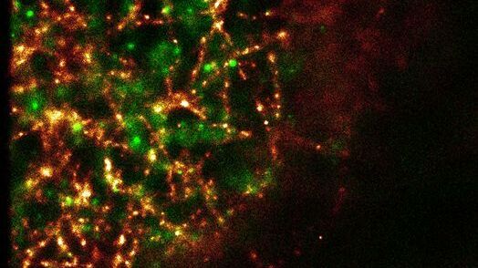
全内反射荧光显微镜(total internal reflection fluorescent microscope,TIRFM)在生命科学研究中的应用
全内反射荧光显微镜的独特之处在于利用衰逝波激发荧光团。与传统的弧光灯、LED 或激光宽场荧光照明方式不同,衰逝波仅能从盖玻片/介质界面开始穿透样本约 100 纳米深度。
Loading...
超分辨率 GSDIM 显微镜
纳米级技术GSDIM(基态耗尽显微镜后单分子返回)提供了细胞内蛋白质和其他生物分子空间排列的详细图像。目前市场上已有首个商业系统(Leica SR GSD),它正在帮助将GSDIM技术推广给更多研究实验室和成像中心的用户。
