
Filter articles
标签
产品
Loading...

荧光和量子点的基本原理和发展历史
在您的科研生涯的某个时候,都有可能会用到荧光显微镜。这种无处不在的技术改变了显微镜学家对研究对象进行成像、标记和追踪的方式,不论是整个生物体,还是单个蛋白质等等。
通过本文,我们将探讨什么是“荧光”,包括其定义背后的历史和基础物理原理,绿色荧光蛋白(GFP)的发现和应用,并展望量子点等荧光探针不断扩大的应用领域。
Loading...
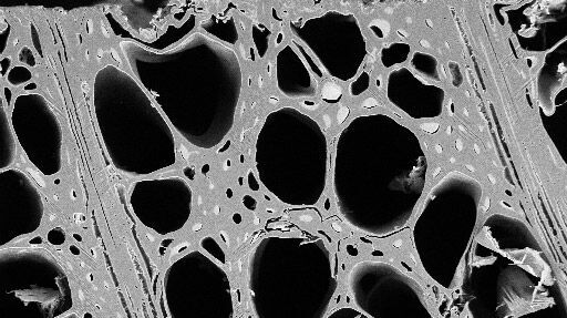
非聚焦离子束研磨的实践应用
机械抛光不仅费时费力,而且还会产生伪影,干扰扫描电子显微镜(SEM)的电子背散射衍射(EBSD)结果或光学显微镜研究。相比之下,离子束研磨可以消除干扰数据分析和图像解读的伪影。
Loading...

不可能的任务:可调颜色用于非扫描检测
徕卡显微系统的 4Tune 探测器是 SP8 DIVE 深度体内探测器的关键组件,提供具有非扫描检测的光谱可调图像记录,是多参数多光子显微镜的创新解决方案。
Loading...

Laser Beam Shaping for Multicolor Multiphoton Microscopy
Multiphoton Microscopy is one of the current hot topics in life science research. The new Leica TCS SP8 DIVE from Leica Microsystems presents a series of beneficial new innovations, including a freely…
Loading...
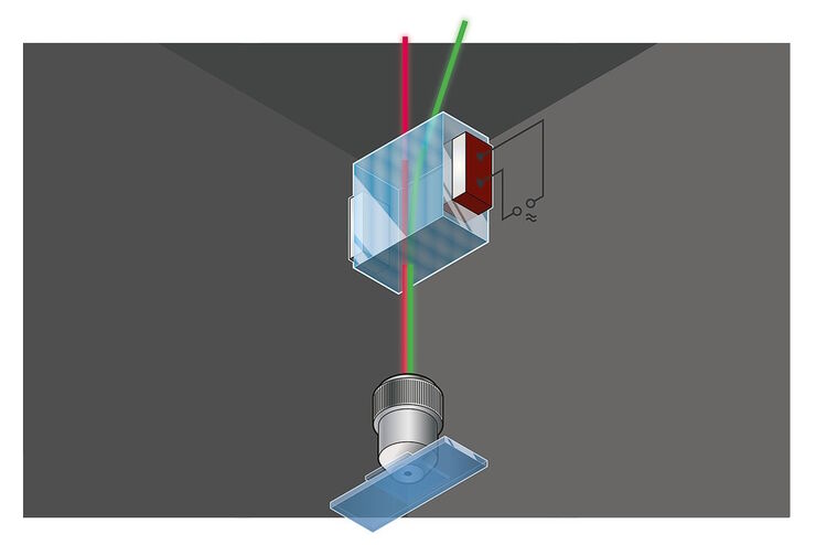
共聚焦显微镜的主分光装置
当前荧光显微镜采用入射照明,这需要将照明和发射光分开。传统的分光设备是一个颜色依赖的分光镜,它具有固定的光谱参数,并通常在指定的波长带内透射90%至98%的发射光。透射率依赖于波长,但也受到技术、设计和实验需求的影响。声光光束分割器(AOBS)则是一种可自由调节的反射缺口设备,平均95%的发射光在这些狭窄的缺口间透射。
Loading...

光激活、光转化和光控开关荧光蛋白
荧光蛋白(FP)如GFP、YFP或DsRed都是可视化观察活细胞中细胞组分的强大工具。尽管如此,目前仍然会有传统FP达到极限的情形发生。普通FP无法观察特定兴趣蛋白中专有、空间有限的蛋白质群体,因为它们在整个细胞中都有表达。此时,光激活、光转化和光控开关荧光蛋白就登上了舞台。荧光套件中的成员可以从非荧光状态激活,可以改变发射光谱,甚至可以逆转式地“开启和关闭”。借助这些“光学荧光笔”,研究人员可以…
Loading...

共聚焦显微镜针孔效应
在操作共聚焦显微镜,或在讨论这种装置的特性和参数时,我们不可避免地提到针孔及其直径。这篇简短的文章是针对那些没有足够时间钻研共聚焦显微镜的理论和细节但又想了解针孔效应的用户们来解释针孔的意义。
Loading...
 stereo microscope for a task like surgery.")
Rodent and Small-Animal Surgery
Learn how you can perform rodent (mouse, rat, hamster) and small-animal surgery efficiently with a microscope for developmental biology and medical research applications by reading this article.
Loading...
 illuminated with wide-band UV excitation. Note the tissue structure with blue autofluorescence. Right: Same tissue and same immunostaining with FITC label illuminated with epi-il")
Milestones in Incident Light Fluorescence Microscopy
Since the middle of the last century, fluorescence microscopy developed into a bio scientific tool with one of the biggest impacts on our understanding of life. Watching cells and proteins with the…
