
Filter articles
标签
产品
Loading...

使用旋转设备进行光片样本安装
TCS SP8 DLS 显微镜采用了一种创新的设计理念,将光片显微技术集成到共聚焦显微镜中。得益于其独特的光学架构,样本可以像进行标准共聚焦成像一样,安装在标准玻璃底培养皿上。与传统的样本安装程序相比,这一过程只需进行少量调整。
Loading...
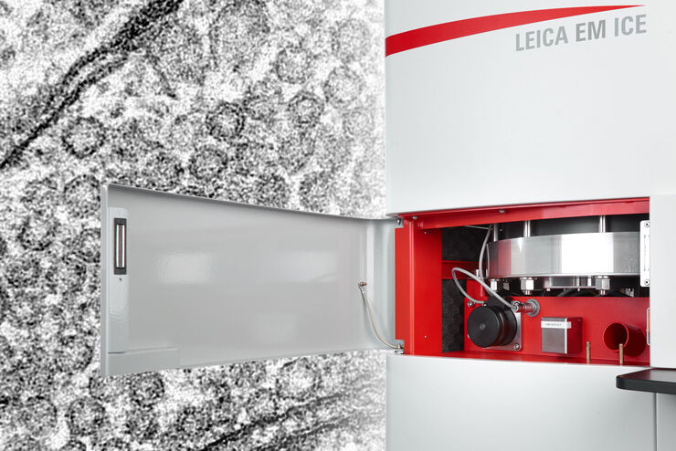
Expanding the Limits of Electron Microscopy Sample Preparation
Capturing the intricate changes in fine structure or in cell dynamics with conventional cryo solutions can be challenging sometimes. Leica Microsystems has developed a new cryo platform, the Leica EM…
Loading...

Live Cell Isolation by Laser Microdissection
Laser microdissection is a tool for the isolation of homogenous cell populations from their native niches in tissues to downstream molecular assays. Beside its routine use for fixed tissue sections,…

Loading...

页岩和碳酸盐岩的宏观至纳米级孔隙分析
页岩和碳酸盐岩等岩石的物理孔隙度对其储存能力有很大影响。孔隙的几何形状也会影响其渗透率。对可见孔隙空间进行成像,有助于了解物理孔隙空间、孔隙几何形状以及与储存和运输相关的矿物和有机物质阶段。

Loading...

DNA分子的可视化
在透射电子显微镜(TEM)中可以使用重金属(如铂)精确的低角度旋转阴影来观察之前在薄的、低晶粒和电子透明的碳膜上吸收的物体的分子细节。
为了达到最高的对比度和更好的图像质量,涂层具有方向性至关重要,它是以精确的角度向样品给出的。金属层的细晶粒和涂层材料在样品表面的均匀厚度也是实现高质量TEM图像的关键要求。这需要电子束蒸发方法,尚无替代涂层技术能够实现相当的结果。电子束蒸发产生的蒸发材料流是非常…
Loading...

使用 U 形玻璃毛细管进行样品装载
徕卡显微系统的DLS显微镜系统是一种创新概念,将光片显微技术集成到共聚焦平台中。由于其独特的光学结构,样本可以安装在标准玻璃底培养皿上,与传统的安装程序相比,几乎不需要或只需很少的适应。在这里,我们介绍了一种便捷的方法,能够快速准备样本以进行光片成像。
