无限远光学系统
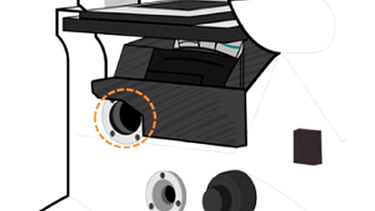
“无限远光学”这一概念是指在显微镜的物镜和镜筒透镜之间具有平行光线的光束路径。平面光学元件可以进入到这个“无限远空间”中,而不影响成像,这对于利用DIC或荧光等对比度方法至关重要。
现代显微技术需要在无限远光路中添加多种光学仪器,如光源或激光装置。满足这一需求的不同方法已经出现,本文对其进行了描述。
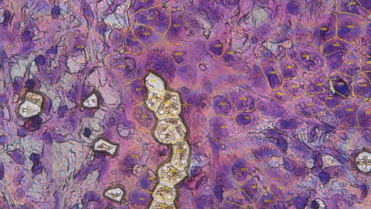
人工智能与深度视觉蛋白质组学 (DVP) 相结合,推进疾病研究
在这次网络研讨会上,Andreas Mund 博士将介绍深度可视蛋白质组学(DVP)--一种将人工智能驱动的组织空间分辨、非靶向蛋白质组学相结合的尖端平台。他展示了 DVP 如何从最小的、表型匹配的细胞群中识别数千种蛋白质,并在复杂的临床组织样本中生成高分辨率分子图谱,从而在细胞水平上解码疾病机制。
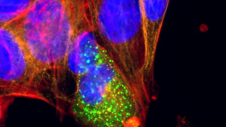
用于二维细胞培养的显微镜和AI解决方案
这本电子书探讨了显微镜和AI技术在二维细胞培养工作流程中的整合。报告重点介绍了明视野、相衬和荧光等传统成像方法如何支持常规细胞监测,而 Mateo TL 和 Mateo FL 数字式倒置显微镜则通过自动汇合检查、细胞计数和转染分析提高了可重复性。它还展示了综合数据管理、审计跟踪和样本跟踪如何改进文档和研究的完整性。本书最后展望了未来趋势,包括微流控技术和 2D-3D…
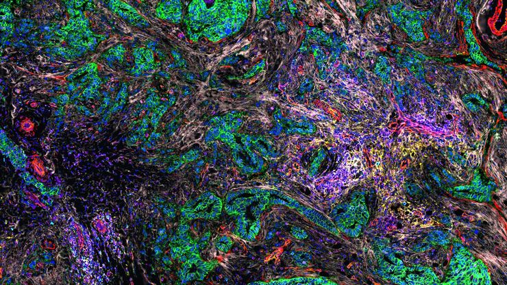
人工智能驱动的乳腺癌研究多重染色成像空间分析工具
乳腺癌(BC)是女性因癌症死亡的主要原因,研究查肿瘤微环境(TME)对于阐明肿瘤进展机制至关重要。利用超多标染色空间蛋白质组学技术系统地绘制肿瘤微环境图谱可以提高精准免疫肿瘤学的能力。在这里,我们将基于人工智能的高倍空间分析应用于BC组织,研究免疫细胞类型和生物标记物,从而深入了解受免疫疗法反应的TME分子机制。
利用光片显微技术聚焦三维长时程成像
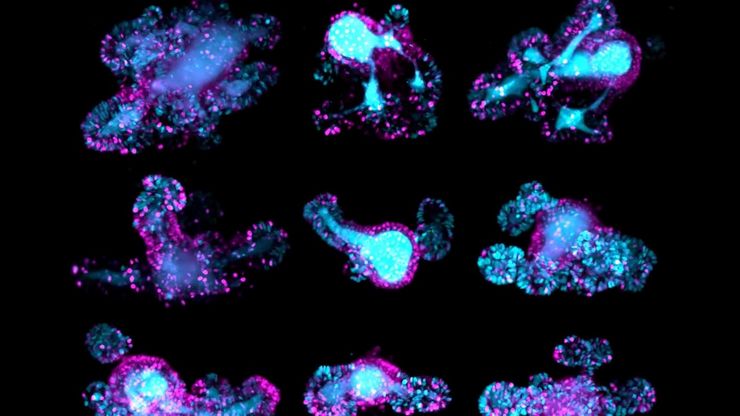
长时程三维成像揭示了复杂的多细胞系统是如何生长和发育的,以及细胞是如何随着时间的推移而移动和相互作用的,从而揭示了发育、疾病和再生方面的重要知识。光片显微镜一次只照射样品的一个薄片,大大减少了光损伤,保护了样品的活性。这种温和的高速技术可在数小时甚至数天内提供清晰的体数据,使研究人员能够实时捕捉生物学的发展过程。
聚合物透射电镜分析用超薄切片技术
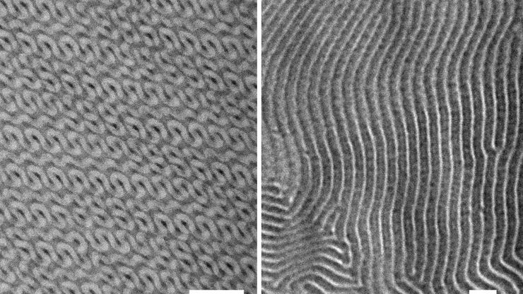
本文全面展示了徕卡UC Enuity超薄切片机在聚合物样品超薄切片制备中的优异表现,无论是常温还是低温环境,它都能提供理想的分析样本。文中展示的高分辨率二维及三维TEM图像,有力印证了该仪器在聚合物结构分析领域,对于获得精确、可重复的样品制备结果不可或缺。
利用激光显微切割发现生物标记物
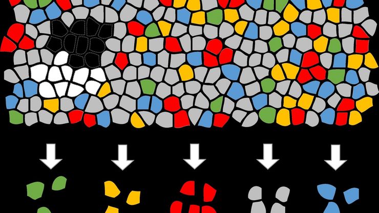
探索空间蛋白质组学工作流程的潜力,如深度视觉蛋白质组学(DVP),以破译病理机制和发现药物靶点。蛋白质表达、丰度或活性的改变会严重影响细胞功能--通常会导致疾病。值得注意的是,相邻细胞之间的蛋白质组可能存在巨大差异。空间蛋白质组学关注到这种细胞异质性,从而揭示了病理机制。激光显微切割技术(LMD)可获取单细胞进行下游分析,同时保留其空间环境,为空间蛋白质组学奠定了基础。
Volume EM and AI Image Analysis
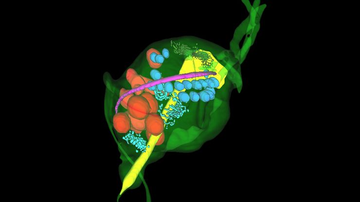
The article outlines a detailed workflow for studying biological tissues in three dimensions using volume-scanning electron microscopy (volume-SEM) combined with AI-assisted image analysis. The focus…
线虫研究指南 - 针对线虫的相关工作
本指南概述了可以高效进行线虫的研究显微镜技术。线虫是一种广泛使用的模式生物,与人类有大约 70% 的基因同源性,是研究发育、神经科学、遗传学和衰老的理想生物。它的透明性和易培育性使其成为一个出色的遗传学模型系统。它可以进行高分辨率成像。主要的实验方法包括挑虫、转基因、荧光筛选、成像和记录。

