
Filter articles
标签
产品

Loading...

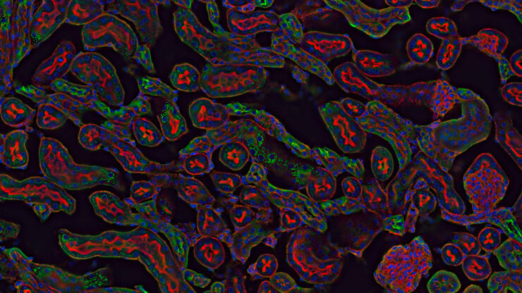
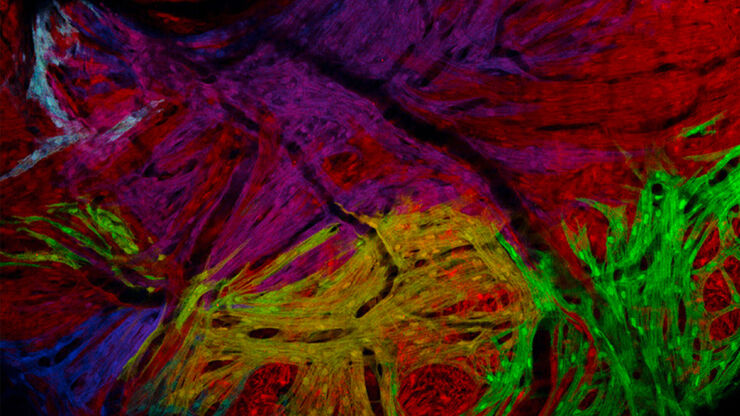
克服显微镜成像移动斑马鱼幼虫时的挑战
Zebrafish is a valuable model organism with many beneficial traits. However, imaging a full organism poses challenges as it is not stationary. Here, this case study shows how zebrafish larvae can be…
Loading...
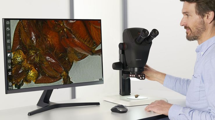
斑马鱼研究
为了在筛选、分拣、操作和成像过程中获取高质量结果,您需要观察细节和结构,从而为您的下一步研究做出正确的决策。
徕卡体视显微镜和透射光底座以出众的光学器件和优良的分辨率而闻名,是全世界研究学者的首选。
Loading...

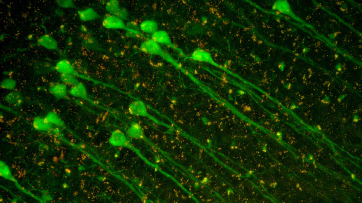
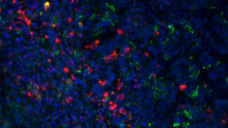
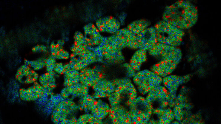
如何在块面中自动获取感兴趣的荧光细胞
本文介绍了使用超薄切片超薄切片机自动修整修块功能,获取树脂块面中带有荧光信号的细胞结构。我们展示了如何使用配置有体视显微镜 M205 FA 的超薄切片超薄切片机 UC Enuity ,来识别感兴趣的荧光细胞,如何自动修整包含细胞的块面,以及如何在切片中观察细胞而无需转移到外部显微镜。
