
 in 14 x 18 tiles. Lifetime gives an additional contrast that allows to differentiate different structures in histological stainings.")
FLIM 方法
虽然绝大多数成像实验都是测量荧光强度,但荧光寿命成像利用荧光的另一个关键特性--荧光寿命--为研究增添了丰富的信息来源。荧光染料具有特征发射光谱和荧光寿命,后者反映了染料在激发态下的停留时间。荧光寿命成像图像的对比度取决于荧光信号的寿命而非强度。由于荧光寿命信息取决于染料的微环境,因此可以为实验提供一些有价值的新见解。
直到最近,对于日常显微镜应用,尤其是涉及活细胞成像的应用来说,荧光寿命成像被认为过于缓慢、复杂和昂贵。但这一切正在迅速改变。凭借当今的先进技术,寿命成像比以往任何时候都更快、更容易使用。
寿命信息与荧光团浓度无关,因此荧光寿命成像在研究分子功能、相互作用和环境方面对功能成像非常有用。生物传感器也可利用荧光寿命成像研究细胞微环境。荧光寿命成像可用于区分具有重叠发射光谱的荧光探针,并消除不必要的背景信号。
在本页中,您可以找到关于荧光寿命成像所有问题的指南,并可链接到相关文章以进一步阅读。了解有关荧光寿命成像基础知识的更多信息。
荧光寿命成像应用文章
A novel concept in fluorescence lifetime imaging enabling video-rate confocal FLIM
Imaging Intracellular Temperature using FLIM


What is FLIM - Fluorescence Lifetime Imaging Microscopy?
基于荧光寿命的成像图库
FLIM 顯微鏡有哪些應用?
FLIM provides information that is not usually available with intensity-only data. Lifetime information can help life science researchers distinguish different fluorophores within a specimen, even if they have overlapping emission spectra. Fluorescence lifetime is independent of molecular concentration, unlike intensity, but it depends on the dye microenvironment, so FLIM allows dynamic changes of local environmental conditions within a specimen to be monitored.
Some popular applications where FLIM can help you gain new insights are:
- Exploring cellular microenvironments with environmentally sensitive fluorescent probes which can be used to track changes in temperature, ion concentration, pH, polarity, viscosity, and secondary messengers.
- Characterization of various types of tissues by autofluorescence.
- Analyzing metabolism and mitochondrial dysfunction in living cells, tissues, and organisms with label-free experiments
Biomedical research applications with FLIM in combination with Förster resonance energy transfer (FRET) has also proven to be beneficial for investigating dynamic changes in cells. FRET allows molecular interactions, like those between ligands and receptors, proteins, or effectors and DNA, to be monitored.
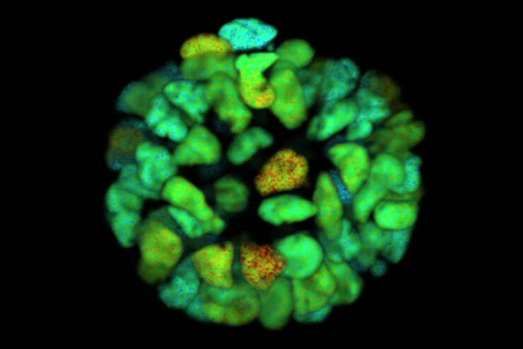
Recent research on diabetes done with FLIM studied the defective maintenance of normal blood glucose levels caused by dysfunction of the alpha and beta cells in the islets. Multiphoton phasor-FLIM NADH autofluorescence imaging was used to detect metabolic changes of living islet cells before and after glucose stimulation. Increased oxidative phosphorylation in beta cells and suppressed oxidative phosphorylation in alpha cells were observed with phasor FLIM after glucose stimulation in healthy islets, but these were not observed in islets with type 2 diabetes.
There are a number of ways that FLIM can add depth to your current confocal experiments, including reducing the chance of artifacts in your image, differentiating the true signal from unwanted autofluorescence, and distinguishing more fluorophores with confidence.
荧光寿命成像应用文章
FLIM相量(Phasor)工具检测代谢差异

Accessing the Metabolic Information of Stem Cells

Structural HIV-1 Env Trimers Dynamics on Living T Cells

为什么荧光寿命成像和佛斯特共振能量转移是如此完美的组合?
荧光是分子或原子在吸收光后激发的光子的自发发射。如果两个荧光分子(荧光团)距离很近,相隔几个纳米,能量就可以直接从 "供体 "转移到 "受体 "荧光团,而不会发生光发射。这种直接的能量交换被称为佛斯特共振能量转移(FRET)。
佛斯特共振能量转移的发生有几种现象。首先,样品(受体)会发出与所应用的激发颜色不同的荧光。这种发射可以测量并与原始发射进行比较;这种方法被称为 "敏化发射"。敏化发射以可量化的方式表明发生了佛斯特共振能量转移。
在另一方面,由于一些激发态转移到受体的激发态,供体的发射将会减少。这种现象被用于一种称为“受体光漂白”的方法中,通过对受体光漂白后供体发射变化的测量来利用这一现象。受体被移除后,供体的发射将会增加。
荧光染料发射光谱的重叠程度越大,发生荧光共振能量转移(FRET)的可能性就越大。即使分子的方向也会影响能量转移。FRET可用于检测和研究细胞中的分子相互作用,例如配体与受体、蛋白质或效应物和核酸之间的相互作用。
基于强度的FRET方法对样本中荧光染料的浓度变化、分子扩散、样本移动以及激发光波动相当敏感。幸运的是,荧光寿命成像(FLIM)与FRET的结合在克服这些限制时提供了巨大的优势。当发生FRET时,供体的荧光寿命会明显减少,甚至可以用作测量FRET效率的一种方法。有关 结合 FLIM 和 FRET 的更多信息。
FLIM 技術最近有哪些進展?
使用 STELLARIS 8 FALCON 系統的快速電子元件和靈敏的光譜混合探測器,可實現具有逐像素量化功能的視頻速率 FLIM。光子到達時間以標準共焦成像的典型計數率記錄。該系統具有超短死區時間和強大的內置演算法,可用於資料擷取和分析。傳統的時間相關單光子計數 (TCSPC) 解決方案本質上速度慢且難以實現,因此 FLIM 成像一直僅限於專家使用,無法提供研究生物過程所需的速度,研究速度超過數十秒。
為了快速獲得結果,壽命成像已經無縫集成到 STELLARIS 8 FALCON 平台的全光譜靈活共焦成像和處理工具中。壽命資訊被記錄為典型共焦影像的額外對比通道。因此,記錄 FLIM 數據現在就像按一個按鈕一樣簡單。
STELLARIS 獨特的 TauSense 技術可讓您從每個樣品中擷取多一層資訊,增加研究的科學影響力。TauSense 由基於螢光生命週期的應用導向成像工具組成,您可以利用這些工具探索分子在細胞環境中的功能:
- TauContrast > 存取新陳代謝狀態、pH 值和離子濃度等功能資訊
- TauGating > 移除不需要的螢光貢獻;以及
- TauSeparation > 擴大光譜選項以外的螢光訊號組合。
- TauInteraction > 直接偵測和量化分子互動 (例如蛋白質與蛋白質互動)。
FLIM FAQ
利用荧光寿命成像可以跟踪细胞环境的动态变化。因此,利用荧光寿命成像可以监测活细胞中蛋白质与配体、受体和核酸等其他生物大分子之间的相互作用。
荧光寿命成像可用于消除标本成像过程中的自发荧光背景。寿命测量可将自发荧光与所需的荧光团信号区分开来。这种能力意味着荧光寿命成像可用于从几乎所有荧光信号(甚至是自发荧光)中收集有意义的信息。
生物医学研究人员通常对监测离子、蛋白质、生物大分子及其在活细胞中的相互作用感兴趣,以了解动态过程。与基于强度的荧光相比,荧光寿命成像是研究细胞动态的更好方法。其寿命与荧光团浓度、照明强度、光吸收和散射无关。


 and acceptor (A) molecule which participate in FRET (Förster resonance energy transfer).")

