

Filter articles
标签
产品
Loading...
斑马鱼研究
为了在筛选、分拣、操作和成像过程中获取高质量结果,您需要观察细节和结构,从而为您的下一步研究做出正确的决策。
徕卡体视显微镜和透射光底座以出众的光学器件和优良的分辨率而闻名,是全世界研究学者的首选。
Loading...

如何在块面中自动获取感兴趣的荧光细胞
本文介绍了使用超薄切片超薄切片机自动修整修块功能,获取树脂块面中带有荧光信号的细胞结构。我们展示了如何使用配置有体视显微镜 M205 FA 的超薄切片超薄切片机 UC Enuity ,来识别感兴趣的荧光细胞,如何自动修整包含细胞的块面,以及如何在切片中观察细胞而无需转移到外部显微镜。
Loading...

组织中的精密空间蛋白质组学信息
尽管可使用基于成像和质谱的方法进行空间蛋白质组学研究,但是图像与单细胞分辨率蛋白丰度测量值的关联仍然是个巨大的挑战。最近引入的一种方法,深层视觉蛋白质组学(DVP),将细胞表型的人工智能图像分析与自动化的单细胞或单核激光显微切割及超高灵敏度的质谱分析结合在了一起。DVP在保留空间背景的同时,将蛋白丰度与复杂的细胞或亚细胞表型关联在一起。
Loading...

How to Study Gene Regulatory Networks in Embryonic Development
Join Dr. Andrea Boni by attending this on-demand webinar to explore how light-sheet microscopy revolutionizes developmental biology. This advanced imaging technique allows for high-speed, volumetric…
Loading...

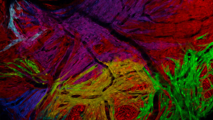
阿尔茨海默病神经免疫相互作用的空间分析
阿尔茨海默病(AD)是一种复杂的神经退行性疾病,以神经原纤维缠结、β-淀粉样斑块和神经炎症为特征。这些功能障碍由局部免疫反应触发或加剧。因此,在空间背景下理解神经免疫相互作用对于阐明 AD 发病机制至关重要。本研究采用 Cell DIVE 多重成像技术和 Aivia 人工智能辅助空间分析工具,探究 AD 病理标志物周围免疫细胞的特征。
Loading...

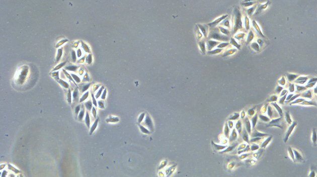
An Introduction to Laser Microdissection
The heterogeneity of histological and biological specimens often requires isolation of specific single cells or cell groups from surrounding tissue before molecular biology analysis can be carried…
Loading...

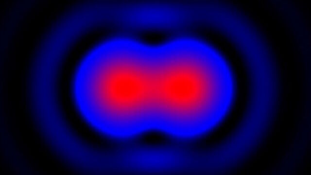
前沿成像技术用于 GPCR 信号传导
通过这个按需网络研讨会,提升您的药理研究,了解 GPCR 信号传导,并探索旨在理解 GPCR 信号如何转化为细胞和生理反应的尖端成像技术。发现领先的研究,扩展我们对这些关键通路的认识,以寻找新的药物发现途径。
Loading...

Revealing Neuronal Migration’s Molecular Secrets
Different approaches can be used to investigate neuronal migration to their niche in the developing brain. In this webinar, experts from The University of Oxford present the microscopy tools and…
Loading...

探索微生物世界:三维食品基质中的空间相互作用
Micalis 研究所是与 INRAE、AgroParisTech 和巴黎萨克雷大学合作的联合研究单位。其使命是开发食品微生物学领域的创新研究,以促进健康。在这一系列视频中,Micalis…
