Filter articles
标签
产品
Loading...

在资源有限的情况下开设神经外科
撒哈拉以南非洲地区的神经外科面临着诸多挑战。在徕卡神经可视化峰会(Leica Neurovisualization Summit)期间,来自世界各地的神经外科医生汇聚一堂,Claire Karekezi 博士在独家网络研讨会上分享了她在资源有限的情况下建立神经外科的经验。
Loading...

Coherent Raman Scattering Microscopy Publication List
CRS (Coherent Raman Scattering) microscopy is an umbrella term for label-free methods that image biological structures by exploiting the characteristic, intrinsic vibrational contrast of their…
Loading...

ISO 9022 标准第 11 部分 - 在苛刻条件下测试显微镜
显微镜和其他光学仪器会受到环境因素的影响。环境因素取决于地理位置和使用地点的条件。仪器的坚固性可以通过完善的加速测试方法进行评估。ISO 9022 标准第 11 部分规定了测试光学仪器抗霉菌和真菌生长的方法。
Loading...

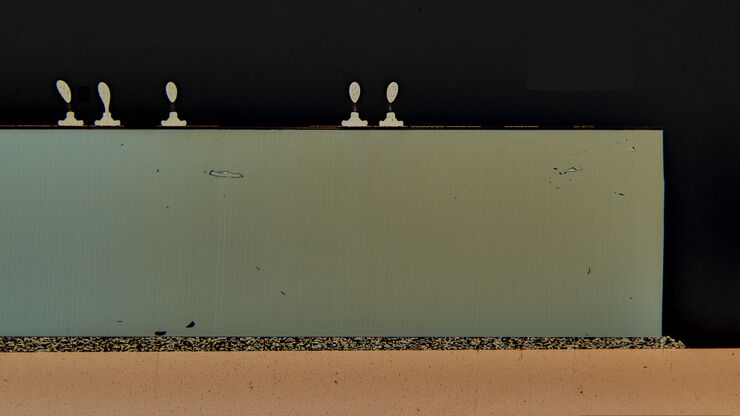
横截面切片法分析IC芯片的结构与化学成分
从本文中了解如何通过横截面分析法对集成电路 (IC) 芯片等电子元件进行有效的结构和元素分析。探索如何通过研磨系统进行铣削、锯切、磨削和抛光工艺以及用于同时进行目视检测和化学分析的二合一解决方案来完成的。可针对电子行业的各种工作流程和应用实现快速、详细的材料分析,包括竞争分析、质量控制 (QC)、故障分析 (FA) 以及研发 (R&D)。
Loading...

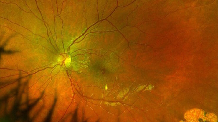
使用光学相干断层扫描改善黄斑孔手术
黄斑裂孔是一种罕见的眼疾,会导致中心视力模糊,影响日常活动。黄斑裂孔通常是由黄斑被牵拉或拉伸而引起的开口,最常见的原因通常是年龄相关的眼部变化。然而,在本病例研究中,Robert A. Sisk博士(医学博士,FACS)介绍了一个小儿眼科病例,其中术中光学相干断层扫描(OCT)为他的手术提供了额外的见解。
Loading...

高质量EBSD样品制备
本文介绍了一种使用宽离子束研磨技术为“混合”晶体材料制备可靠且有效的EBSD(电子背散射衍射)样品的方法。该方法产生的横截面具有高质量表面,这对于EBSD分析至关重要。电子背散射衍射(EBSD)材料分析是通过扫描电子显微镜(SEM)进行的。制备混合材料(CPU或铝(Al)、金刚石和石墨(C)的复合材料)的横截面,使其具有适合EBSD分析的高质量表面,可能是一个挑战。
Loading...

How Marine Microorganism Analysis can be Improved with High-pressure Freezing
In this application example we showcase the use of EM-Sample preparation with high pressure freezing, freeze substiturion and ultramicrotomy for marine biology focusing on ultrastructural analysis of…
Loading...

AR荧光在神经血管手术中的应用
术中血管造影在神经血管手术中发挥着至关重要的作用。在徕卡2021神经可视化峰会期间,Christof Renner博士在独家网络研讨会上展示了精选临床病例,并分享了使用GLOW800增强现实荧光技术的经验。
Loading...

生命科学研究: 哪种显微镜相机适合您?
相机是显微镜系统的重要组成部分,对系统的性能有重大影响。在选择相机时,重要的是不仅要看技术规格,还要考虑您的样品、技术、对比方法以及您希望获得的数据类型。

