工业

工业

工业

深入探讨有关工业和病理学领域的高效检测、优化工作流程和提高人体工学舒适度的文章和网络研讨会。涉及的主题包括质量控制、材料分析、病理学显微镜等。在这里您可以获得有关使用前沿技术提高生产力和优化质量以及准确地进行病理学诊断的干货。
[Translate to chinese:] Area of a printed circuit board (PCB) which was imaged with extended depth of field (EDOF) using digital microscopy.

如何形成清晰的图像

在显微镜检查中,景深常被看做经验参数。在实际操作中,会根据数值孔径、分辨率和放大率之间的相关性确定该参数。为了获得最佳视觉效果,现代显微镜的调节设备在景深和分辨率之间实现了最佳平衡,这两个参数在理论上呈负相关。
[Translate to chinese:] These images show the microstructure of a hard metal with 10% cobalt which is used for heavy-duty tools. The large increase in magnification of the right image (compared to the left) has a risk of being outside the useful range or, in other words, empty magnification.

当心“无效”放大率

在一个最简化的情况,光学显微镜由一个靠近标本的透镜(物镜)和一个靠近眼睛的透镜(目镜)组成。显微镜放大率是两个显微镜透镜系数的乘积。比如,40倍物镜和10倍目镜可以得到400倍放大率。
[Translate to chinese:] Multicolor TauSTED Xtend 775 for Cell Biology applications that require nanoscopy resolution for multiple cellular components. Cells showing vimentin fibrils (AF 594), actin network (ATTO 647N), and nuclear pore basket (CF 680R). Sample courtesy of Brigitte Bergner, Mariano Gonzales Pisfil, Steffen Dietzel, Core Facility Bioimaging, Biomedical Center, Ludwig-Maximilians-University, Munich, Germany.

STED样品制备指南

这份指南旨在帮助用户优化受激发射损耗(STED)纳米成像的样品制备,特别是在使用徕卡微系统的STED显微镜时。它提供了单色STED成像用荧光标记的概述,并对其性能进行了评级。
An 8-color spectral unmixing result from a hyperspectral SRS (stimulated Raman scattering) dataset, showing the biochemically distinct structures of a fresh, untreated apple slice.

How to Prepare Samples for Stimulated Raman Scattering (SRS) imaging

Find here guidelines for how to prepare samples for stimulated Raman scattering (SRS), acquire images, analyze data, and develop suitable workflows. SRS spectroscopic imaging is also known as SRS…
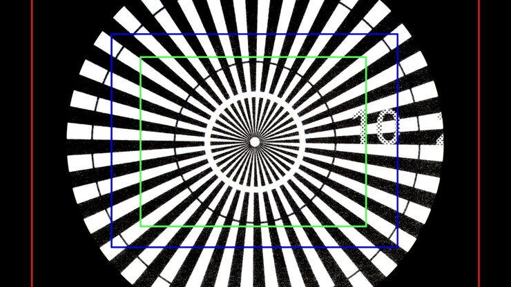
Image of a Siemens star, where the diameter of the 1st black line circle is 10 mm and the 2nd is 20 mm, taken via an eyepiece of a M205 A stereo microscope. The rectangles represent the field of view (FOV) of a Leica digital camera when installed with various C-mounts (red 0.32x, blue 0.5x, green 0.63x).

30000:1放大率到底意味着什么?

关于光学显微镜性能的一个重要标准是放大率。本报告将为数字显微镜用户提供有用的指南,以确定放大率值的有用范围。
[Translate to chinese:] Molecular structure of the green fluorescent protein (GFP)

荧光蛋白简介

本文概述了荧光蛋白及其光谱特性。随着 20 世纪 50 年代末荧光蛋白的发现,荧光显微技术发生了巨大变化。它始于 O. Shimomura 和来自水母(Aequorea victoria)的绿色荧光蛋白(GFP)[1]。后来出现了数百种 GFP…
Micrograph of dinoflagellate cells. Scale bar = 1 µm.

How Marine Microorganism Analysis can be Improved with High-pressure Freezing

In this application example we showcase the use of EM-Sample preparation with high pressure freezing, freeze substiturion and ultramicrotomy for marine biology focusing on ultrastructural analysis of…
Patch pipette touching a murine hippocampal neuron. Image courtesy of A. Aguado, Ruhr University Bochum, Germany.

什么是膜片钳技术?

离子通道的生理学一直是神经科学家感兴趣的一个重要话题。诞生于1970年代的膜片钳技术开启了电生理学家的新时代。它不仅可以对整个细胞进行高分辨率电流记录,还可以对切下的细胞膜片进行高分辨率电流记录。甚至可以研究单通道事件。然而,由于需要复杂且高灵敏的设备,广泛的生物学背景和高水平的实验技能,电生理学仍然是最具挑战性的实验室方法之一。
[Translate to chinese:] Neurons imaged with DIC contrast.

微分干涉对比(DIC)显微镜

本文阐释了在使用显微镜对未染色透明生物样本进行成像时,微分干涉对比法(DIC)为何是明场照明的绝佳方案。
Scroll to top