
Filter articles
标签
产品
Loading...

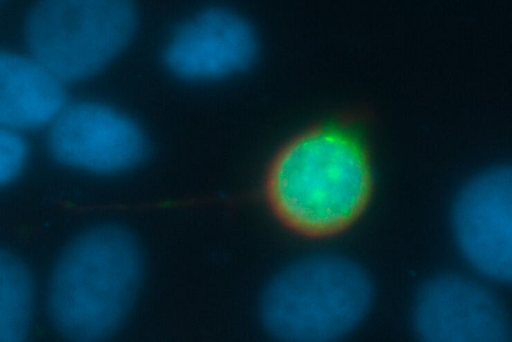
人工智能显微成像能够高效检测稀有事件
对稀有事件进行定位和选择性成像是许多生物样本研究过程的关键。然而,由于时间限制和高度的复杂性,有些实验无法做到,从而限制了获得新发现的前景。通过基于人工智能的显微成像检测稀有事件,这种工作流程将智能样本导航、图像采集工具和人工智能驱动的图像分析等不同功能融合起来共同协作,能够克服上述局限性。
Loading...

荧光入门介绍
荧光是George Gabriel Stokes于1852年首次报道的一种现象。他观察到萤石在紫外线照射后开始发光。荧光是光致发光的一种形式,是指一种材料被光照射后会发射出光子。发射光的波长比激发光更长。这种效应又称为斯托克斯位移。

Loading...

借助多重成像深入了解胰腺癌的复杂性
胰腺癌是一种很难治疗的肿瘤疾病。Cell DIVE多重成像可以视觉呈现30种生物标志物以探测胰管癌的微环境。此面板可以检查肿瘤组织多个层级的问题,包括淋巴细胞、血管新生、转移、侵袭、炎症、缺氧、代谢和免疫。多重成像和分析可以对肿瘤组织中的许多生物活动提供更为深入的洞察信息,从而可以深入研究这些信息。


Loading...

什么是膜片钳技术?
离子通道的生理学一直是神经科学家感兴趣的一个重要话题。诞生于1970年代的膜片钳技术开启了电生理学家的新时代。它不仅可以对整个细胞进行高分辨率电流记录,还可以对切下的细胞膜片进行高分辨率电流记录。甚至可以研究单通道事件。然而,由于需要复杂且高灵敏的设备,广泛的生物学背景和高水平的实验技能,电生理学仍然是最具挑战性的实验室方法之一。
Loading...

如何利用单个系统对组织学荧光样品进行成像
在本集MicaCame中,主持人Lynne Turnbull和Patric Pelzer将带您探寻生物样本染色的历史之旅。他们将解释为什么您通常必须选择为组织学样品或荧光样品选择特定的系统,以及如何利用新的成像技术克服这一点。
Loading...
超越反卷积
宽场荧光显微镜通常用于视觉呈现生命科学样本中的结构并获取重要信息。利用荧光蛋白或染料,以高度特异性的方式标记离散的样本部分。为了充分了解某种结构,可能需要以三维方式呈现,但这会对使用显微镜带来某些挑战。
