Filter articles
标签
产品
Loading...
 acquired using THUNDER Imager Live Cell. Image courtesy of Janina Kaspar and Irene Santisteban, Schäfer Lab, TUM.")
研究大脑健康的成像类器官模型
小胶质细胞是特化的脑驻留免疫细胞,在大脑发育、平衡和疾病中发挥着至关重要的作用。然而,到目前为止,模拟人脑环境与小胶质细胞之间相互作用的能力还非常有限。
. Courtesy: Thomas Mathivet, PhD")
Loading...

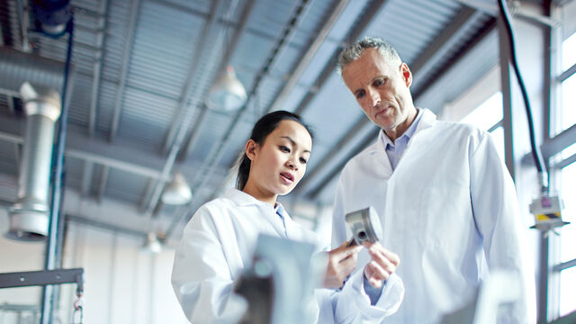
如何使显微镜工作场所符合人体工学
对于每天使用显微镜工具的人来说,显微镜具有重要影响。显微镜对人体有着很高的要求,需要我们集中注意力,运用肌肉从事大量稳定活动。
在此次采访中,Leica Microsystems的高级产品经理Clinton Smith谈到了如何缓解可能出现的紧张和压力,如何创造符合人体工学的工作场所来帮助显微镜用户舒适地工作,以及如何提高生产率。
Loading...
 and labelled with MitoTracker Green.")
可重复性、协作和新成像技术的力量
在本次网络研讨会上,您将了解到影响显微镜可重复性的因素,有哪些资源和举措可用于改善显微镜教育并提高其严谨性和可重复性以及研究人员、成像科学家和显微镜供应商之间的合作如何推动创新和采用新技术。
Loading...

检查癌症类器官的发展进程
德国慕尼黑工业大学的Andreas Bausch实验室研究细胞和生物体中不同结构和功能形成的细胞和生物物理机制。他的团队设计了新的策略、方法和分析工具,以量化微米和纳米等级的发展机制和动态过程。关键研究领域包括干细胞和类器官,从乳腺类器官到胰腺癌类器官,以更好地了解疾病模型。
. Image courtesy of Prof. Hui Guo, School of Life Sciences, Central South University, China")
Loading...

借助人工智能,揭示复杂而密集的神经元图像中的洞察
神经元的3D形态学分析通常需要使用不同的成像模式,捕捉多种类型的神经元,并在各种密度下相连的传统Leica SP8显微镜采集多达解神经元的形态,这对许多研究人员来说仍然是一个耗时的挑战。
Loading...
 tissue on a single slide.")
表征肿瘤环境以揭示洞察和空间分辨率
肿瘤环境的表征可以为癌症进展和潜在治疗靶点提供更深入的见解。我们已经使用来自Cell Signaling Technology(CST)的各种IHC验证抗体,在胰腺癌的Cell DIVE研究中验证了30多种偶联抗体。
Loading...

神经科学显微镜面临哪些挑战?
显微镜是神经科学研究领域的强大工具。不过,当涉及到对神经过程进行成像以及使用不同的样品类型(例如厚神经组织或脑类器官)时,科研人员可能会面临到很多挑战。这本30页的电子书包含众多真实的案例,以讨论我们最常见到的一些挑战,同时展示了如何使用THUNDER 成像技术克服这些挑战。
