
Filter articles
标签
产品
Loading...
加速不同组织多重成像的发现
组织的多重成像对于肿瘤-免疫相互作用的研究以及人类细胞图谱等发现工作越来越重要。 欢迎加入我们的演讲,Andrea J. Radtke 博士解释了如何使用迭代漂白扩展多重性 (IBEX) 绘制组织图谱,并讨论了用于多重成像的广泛社区资源。

Loading...

基于 AI 引导的多重二维数据向空间洞察的转化
Aivia 13 能够处理大型二维图像,使研究人员能够通过检测数百万个对象和自动聚类多达 30 个标记物,深入理解其表型周围的微环境。


 imaged with the THUNDER Imager 3D Cell Culture. Courtesy of Dr. F.T. Arroso Martins, Tamere University, Finland.")
Loading...

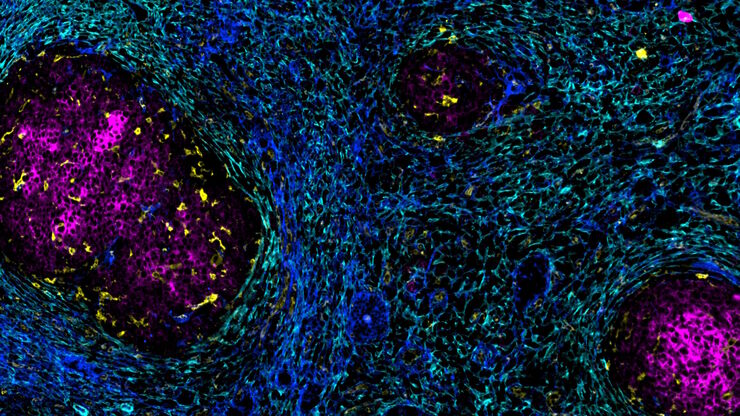
肝细胞癌中癌症干细胞位点的原位鉴定
在这里,我们探索了一种突破性的多重免疫检测方法,通过多重成像对细胞外基质(ECM)特征进行原位定位,从而识别肝细胞癌(HCC)内的癌症干细胞龛。

Loading...

落射荧光显微镜和反射对比显微镜
多年来,荧光显微镜一直仅使用透射光和暗场照明。随着时间的推移,对改进照明的需求不断增长,这导致了落射照明(也称为入射光照明)的发展。经过 40 年的发展和改进,落射照明荧光显微镜已成为生命科学、临床医学诊断和材料科学领域常规实验室工作和研究的实用方法。大部分开发工作由 Ploem 集团和 Leitz 公司(现为 Leica Microsystems)完成。
