
Filter articles
标签
产品
Loading...

20 Years of Leica Laser Microdissection
Phenotype-genotype correlations are key for insight. From Eye to Insight is therefore fitting perfectly to Leica Microsystems and in particular to laser microdissection. Laser Microdissection, also…

Loading...

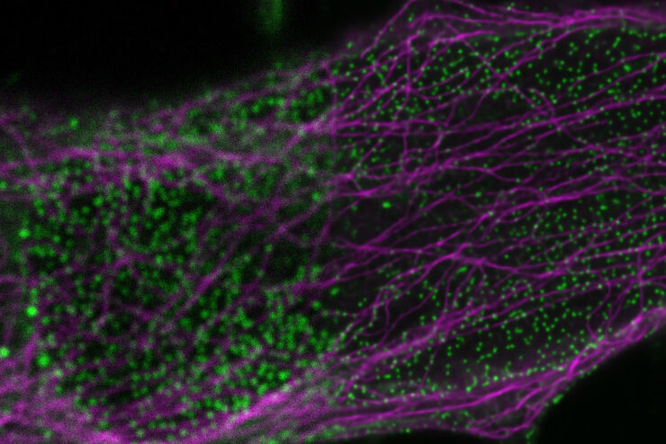
Regulators of Actin Cytoskeletal Regulation and Cell Migration in Human NK Cells
Dr. Mace will describe new advances in our understanding of the regulation of human NK cell actin cytoskeletal remodeling in cell migration and immune synapse formation derived from confocal and…
Loading...

Adding Dimensions to Multiplex Molecular Imaging
Molecular imaging of living specimens offers a means to draw upon the growing body of high-throughput molecular data to better understand the underlying cellular and molecular mechanisms of complex…
Loading...
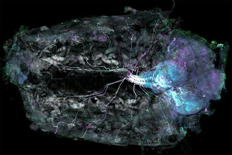
Understanding Motor Sequence Generation Across Spatiotemporal Scales
We have developed a microscopy-based pipeline to characterize a developmentally critical behavior at the pupal stage of development, called the ecdysis sequence. We study brain-wide neuronal activity…
Loading...

TauContrast 对图像复杂样本的好处
在这次访谈中,Timo Zimmermann博士谈到了他在应用TauSense工具方面的经验,以及这些工具在研究厚样本或超大胚胎等复杂样本中的潜力。作为巴塞罗那基因组调控中心(CRG)先进光学显微镜单位的负责人,Timo Zimmermann博士在2020年测试了STELLARIS 5共聚焦系统。
Loading...

使用冷冻共聚焦显微镜定位活性循环核孔复合物
本文介绍了如何利用冷冻光学显微镜,尤其是冷冻共焦显微镜来提高冷冻工作流程的可靠性。评估了EM网格和样品的质量,并分析了目标结构的分布。本文展示了如何将冷冻共焦3D数据投射到SEM图像上,将感兴趣结构可靠地保留在FIB切割的薄片内,以便在冷冻TEM中进行进一步研究。
Loading...

使用增强功能电子显微镜研究大脑切片中的突触
神经科学的一个基本问题就是突触的结构与其功能特性之间有何关系?过去几十年,电生理学揭示了突触传递机制,而电子显微镜(EM)深入探索了突触形态。用于关联突触生理学和超微结构的方法可以追溯到20世纪中叶。目标是获得突触传递的快照,即捕获电子显微照片中的动态过程。
