Filter articles
标签
产品
, TL DIC")
Loading...

Dissecting Proteomic Heterogeneity of the Tumor Microenvironment
This lecture will highlight cutting edge applications in applying laser microdissection and microscaled quantitative proteomics and phosphoproteomics to uncover exquisite intra- and inter-tumor…
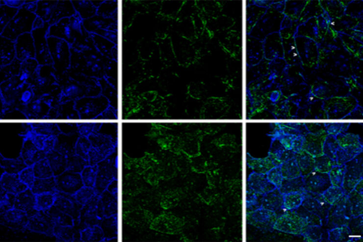
 and astrocytes (green) in a cortical spheroid derived from human induced pluripotent stem cells.")
Loading...

自适应反卷积与 Computational Clearing 结合的力量
反卷积是一种计算方法,用于恢复被点扩散函数(PSF)和噪声源破坏的物体图像。在本技术简介中,您将了解徕卡显微系统提供的反卷积算法如何帮助您克服宽视场 (WF) 荧光显微镜中由于光的波动性和光学元件对光的衍射而造成的图像分辨率和对比度损失。探索由用户控制或自动反卷积的方法,查看并解析更多的结构细节。
Loading...
改进成像技术以了解细胞器膜细胞动态
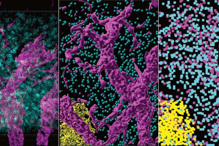
了解正常组织和肿瘤组织中的细胞功能,是推动潜在治疗策略研究和了解某些治疗失败原因的关键因素。单细胞分析在生物医学研究中至关重要,它能揭示在癌症等复杂疾病中哪些细胞和分子通路发生了改变。
Loading...
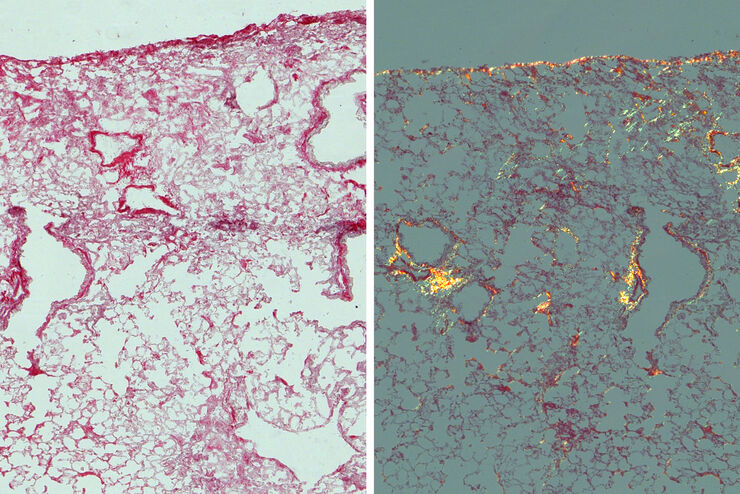
肺纤维化研究
本文中所示结果表明,相比明场,使用偏振光可以更清晰地分辨小鼠肺组织中的胶原蛋白纤维化和非纤维化区域。为更好地理解促使瘢痕组织形成的肺纤维化,正常情况下会研究组织中的纤维化区域。分别使用明场和偏振光对小鼠肺组织中的胶原蛋白纤维化病变区域进行成像。成像胶原蛋白时,使用一般的染色法和明场显微镜很难区分纤维化和非纤维化区域。本文中使用偏振光对肺组织进行成像,两个区域呈现出非常明显的颜色差异。
Loading...

Image Gallery: THUNDER Imager
To help you answer important scientific questions, THUNDER Imagers eliminate the out-of-focus blur that clouds the view of thick samples when using camera-based fluorescence microscopes. They achieve…
Loading...
从器官到组织再到细胞:使用宽场显微镜分析 3D 标本
在传统的宽场显微镜下从厚的三维样本中获取高质量的数据和图像是具有挑战性的,因为存在失焦光的干扰。在本次网络研讨会中,Falco Krüger 展示了THUNDER成像仪如何通过Computational Clearing技术使这一切成为可能。
Loading...

An Introduction to Computational Clearing
Many software packages include background subtraction algorithms to enhance the contrast of features in the image by reducing background noise. The most common methods used to remove background noise…
