
Filter articles
标签
产品
Loading...

How does Real-Time OCT Imaging Impact Precision in Corneal Surgery?
Corneal surgery is a highly specialized field. It requires great surgical precision to overcome challenges such as visualizing clearly the full anterior chamber, performing Descemet membrane peeling…
Loading...

How to Achieve Brain Tissue Resection with GLOW400 AR
Intraoperative MRI is one form of real-time intraoperative visualization, but if more in-depth visualization to identify a tumor during surgery is wanted, intraoperative fluorescence diagnostics is…
Loading...

如何在块面中自动获取感兴趣的荧光细胞
本文介绍了使用超薄切片超薄切片机自动修整修块功能,获取树脂块面中带有荧光信号的细胞结构。我们展示了如何使用配置有体视显微镜 M205 FA 的超薄切片超薄切片机 UC Enuity ,来识别感兴趣的荧光细胞,如何自动修整包含细胞的块面,以及如何在切片中观察细胞而无需转移到外部显微镜。
Loading...

组织中的精密空间蛋白质组学信息
尽管可使用基于成像和质谱的方法进行空间蛋白质组学研究,但是图像与单细胞分辨率蛋白丰度测量值的关联仍然是个巨大的挑战。最近引入的一种方法,深层视觉蛋白质组学(DVP),将细胞表型的人工智能图像分析与自动化的单细胞或单核激光显微切割及超高灵敏度的质谱分析结合在了一起。DVP在保留空间背景的同时,将蛋白丰度与复杂的细胞或亚细胞表型关联在一起。
Loading...

当心“无效”放大率
在一个最简化的情况,光学显微镜由一个靠近标本的透镜(物镜)和一个靠近眼睛的透镜(目镜)组成。显微镜放大率是两个显微镜透镜系数的乘积。比如,40倍物镜和10倍目镜可以得到400倍放大率。
Loading...

How to Study Gene Regulatory Networks in Embryonic Development
Join Dr. Andrea Boni by attending this on-demand webinar to explore how light-sheet microscopy revolutionizes developmental biology. This advanced imaging technique allows for high-speed, volumetric…
Loading...

Spatial Analysis of Neuroimmune Interactions in Alzheimer’s Disease
Alzheimer’s disease (AD) is a complex neurodegenerative disorder characterized by neurofibrillary tangles, β-amyloid plaques, and neuroinflammation. These dysfunctions trigger or are exacerbated by…
Loading...
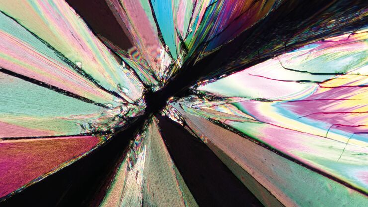
偏振光显微观察
偏光显微镜通常应用于材料科学和地质学领域,根据矿物的折射特性和颜色来识别矿物。在生物学中,偏光显微镜通常用于晶体等双折射结构的识别或成像,或用于植物细胞壁中纤维素和淀粉粒的成像。
Loading...

A Guide to Spatial Biology
What is spatial biology, and how can researchers leverage its tools to meet the growing demands of biological questions in the post-omics era? This article provides a brief overview of spatial biology…
