Explore the features of the THUNDER Imager Live Cell and 3D Assay
THUNDER Imager
Show details
Explore the features of the THUNDER Imager Live Cell and 3D Assay with our interactive image map
Click on the + buttons to experience the technical highlights that support your fluorescence microscopy applications with high resolution imaging and optimal physiological conditions for your live cell cultures.
1
THUNDER Imager 3D Assay
Show details
2
THUNDER Imager Live Cell with Incubator
Show details
3
THUNDER Imager Live Cell
Show details
THUNDER Imager Live Cell was for me the perfect solution to get very high-quality images and also spend little time because it's just so fast to acquire each individual slide. By using THUNDER’s Large Volume Computational Clearing, I was able to remove this typical widefield haze and distinguish between background and signal. And that gave me the perfect starting point for my downstream analysis.
达到高通量,获得更好的统计数据和工作流程效率
为您的 3D 细胞培养试验实现自动化,高效研究新一代疾病模型。THUNDER 能助您对肺器官等大体积样品进行高速成像。此外,自动化还能在繁琐的实验中将用户的操作步骤减至最低。
For 3D cell culture, observing the true physiology is paramount for meaningful results. Typically, you want to investigate your cells while they are in a close to natural state by optimizing experimental conditions, e.g., the lowest light intensity and shortest exposure times possible.
THUNDER Imager Live Cell meets these demands with a high-end LED source that has a small bandwidth optimized for excitation. Even with low illumination and short exposure times, the sensitive high-end sCMOS camera delivers meaningful image information thanks to a quantum efficiency of up to 82%.
To further reduce exposure of the sample to light, illumination is limited to the actual recording time. The camera shutter is synchronized with the high-speed (20 µs switching time) Lumencor LED light source to minimize photobleaching.
[Translate to chinese:] THUNDER Imager Live Cell is based on a fully motorized DMi8 microscope, Quantum Stage, highly sensitive K8 camera, and multi-line, high-intensity fluorescence LED light source. It is optimized for fast and precise multi-position, multi-channel imaging of 3D cell cultures.
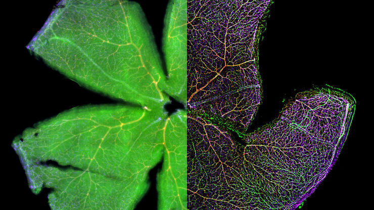
视网膜成像的定量分析方法通常注重于提供视网膜形态和功能的综合描述。视网膜异常以及转化临床应用都需要可靠的工作流程来重现转基因靶点筛选。因此,形态学的重复成像需要能够持续重现准确结果的系统解决方案。使用 THUNDER Imager 3D Assay,您可以清晰地观察形态以及可靠地计算细胞内部细节,例如视网膜中的单个细胞核分布。
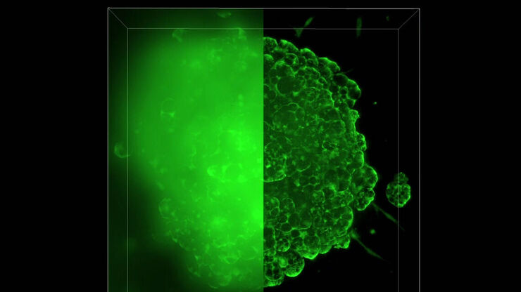
作为新型模式系统,大脑类器官可用于研究人类大脑的发育和疾病。这些自组装式三维细胞结构通常通过多重转基因标记物成像进行表征。这些工作流程中的典型挑战是及时量化分子动力学,同时保持生理条件并在低信号水平下依然能达到样本深度。因此,THUNDER Imager Live Cell适合用于研究接近生理条件的类器官的发育,因为我们的 LED 光源有助于最大限度地减少光漂白。此外,即使蛋白质信号水平低,也可以定时检测而无需改变样本载体。
外植体细胞培养通常难以进行成像实验,因为它们需要稳定的细胞培养环境和低光毒性的成像条件。美国弗吉尼亚大学 Laura Shankman 博士的外植体细胞培养成像实例显示了腹主动脉细胞如何在48小时内稳定成像。THUNDER Imager Live Cell 为微创和活细胞准确成像实验提供完整的显微镜成像系统。凭借快速的高量子效率相机选项、准确的载物台、可调 LED 光源、减少宽场图像中离焦模糊现象的计算清除技术(Computational Clearing)以及易于使用的 LAS X 软件进行自动成像和分析工作流程,可以高效地执行敏感的细胞培养实验。
THUNDER Imager 3D Live Cell 和 THUNDER Imager 3D Cell Culture 能够一次完成全帧摄取,让您体验到高度灵敏、基于 sCMOS 摄像头的荧光系统的强大实力。
结合其高度灵敏性,THUNDER Imager 3D Live Cell 和 THUNDER Imager 3D Cell Culture 可实现高达 90 帧/秒的数据摄取速度,助您观察到快速的细胞活动。即使深入较厚的 3D 细胞团,它也能快速摄取清晰的图像数据。得益于可快速切换的外部滤色片转盘 (< 27 ms),即使在多发射波长的实验过程中,您也能始终掌握快速成像过程。


")

![[Translate to chinese:] THUNDER Imager Live Cell is based on a fully motorized DMi8 microscope, Quantum Stage, highly sensitive K8 camera, and multi-line, high-intensity fluorescence LED light source. It is optimized for fast and precise multi-position, multi-channel imaging of 3D cell cultures.](/fileadmin/_processed_/2/5/csm_THUNDER_Imager_Live_Cell_K8_ThunderShooting-1070585_web_57f5dcf604.jpg)