联系我们

超薄切片机和冷冻超薄切片机

无论是组织样本、聚合物、橡胶、金属或纳米颗粒,徕卡超薄切片机都能提供极薄片以及无缺陷的表面质量。

从材料科学到癌症研究,我们的超薄切片机在全世界被用于各种各样的研究和质量控制。

超薄切片和无缺陷的表面质量!

联系我们

联系当地专家,获取有关符合您需求和预算的专家建议

不妥协的人体工学

用户经常长时间使用超薄切片机。因此,无论是右手使用者还是左手使用者,都必须进行无疲劳的操作。符合人体工程学的扶手和丰富的调整方案使得来自徕卡显微系统的超薄切片机有更舒适的工作体验。

纳米级精度

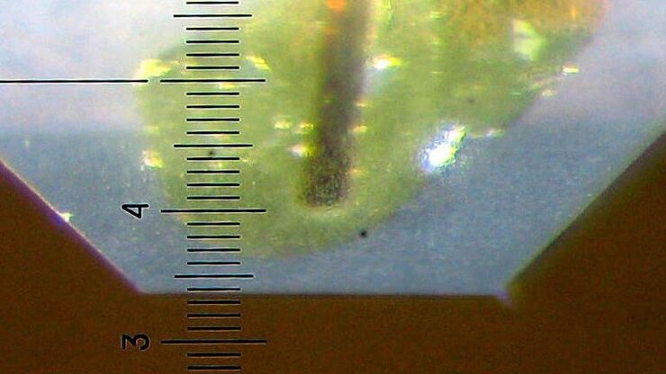
徕卡超薄切片机保证精度和舒适度。由于全电动刀和丰富的技术,甚至初学者都可以准备无缺陷的切片。几分钟内用徕卡EM KMR3为完美的超薄切片做出无缺陷的玻璃刀。

此切片机产生的截面厚度在10 nm到15µm。请探索徕卡 超薄切片机的精密机械并享受为LM、TEM、SEM和AFM检测做出的高质量的样品制备。

相关文章

阅读我们的最新文章

作为徕卡显微系统有限公司 的信息门户,ScienceLab (徕卡课堂) 提供许多关于显微技术主题的科研和教学材料。其内容宗旨在于为初学者、有经验的从业者和科学家等的日常工作和实验提供支持。

更多文章
Block-face created by automatic trimming under fluorescence. Mammalian cells of interest, stained with CellTrackerTM Green are visualized within the block-face using the UC Enuity equipped with the stereo microscope M205 FA. In the background a carbon finder grid in black is visible. All samples in the article are created by Felix Gaedke, PhD, CECAD, Cologne, Germany.

How to Automatically Obtain Fluorescent Cells of Interest in a Block-face

Block-face created by automatic trimming under fluorescence. Mammalian cells of interest, stained with CellTrackerTM Green are visualized within the block-face using the UC Enuity equipped with the…
[Translate to chinese:] UC Enuity

通过自动切片改善您的超薄切片工作流程

在不断发展的电镜样品制备领域,保持领先地位至关重要。这个网络研讨会提供了关于超薄切片最新进展的重要见解,这些进展可以显著增强您实验室的能力。
[Translate to chinese:] Camera image during auto alignment. The feedback lines indicate if the correct edges in the image are detected. Green: Vertical center line; Magenta: Upper edge of the light gap; White: Lower edge of the light gap (not visible here, falling together with red line); Red: Knife edge; Blue: Left and right edge of the block face being automatically detected.

高质量超薄切片:样品与切片刀自动对齐

超薄切片技术是获取样品切片的最常用方法。在室温条件制备时,将样品小块嵌入环氧树脂中,然后通过修剪去除多余的树脂,并使用玻璃刀或金刚石刀将样品切成厚度为50-100纳米之间的薄片。
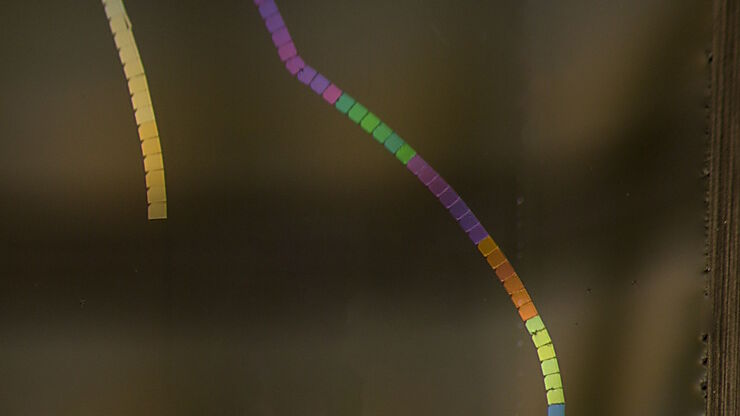
[Translate to chinese:] Section ribbons with increasing section thickness - silver to purple ending in blue sections.

超薄获得高质量的超薄切片

UC Enuity能够应对这一挑战,提供厚度一致且高质量的切片。其用户友好的设计不仅简化了研究过程,还提高了可重复性,使研究者对所得到的结论更有信心。
[Translate to chinese:] Water Flea Daphnia imaged by Electron Microscopy. Courtesy of Mag. Dr. Gruber, University of Vienna, Austria

简化生物应用的电镜样品制备工作流程

电子显微镜(EM)一直处于超微分辨率生物成像的最前沿,它简化了生物应用的 电镜 样品制备工作流程。
[Translate to chinese:] Acrylonitrile butadiene styrene (ABS) stained with osmium tetroxide (OsO4), sectioned with a DIATOME diamond knife at room temperature, and then imaged with HAADF TEM.

用于材料切片的超薄切片技术

了解这些用于材料切片的超薄切片技术的宝贵见解,并学习如何克服在处理聚合物、金属和脆性材料时遇到的挑战。千万不要错过这个提高您对先进显微镜样品制备方法的认识的机会。
Ultramicrotomy; ARTOS 3D

超薄切片介绍

对样本开展研究时,为了以纳米级分辨率显示其精细结构,通常会使用到电子显微镜。电子显微镜有两种类型:扫描电子显微镜(SEM)用于对样本表面成像,以及需要使用极薄电子透明样本的透射电子显微镜(TEM)。因此,使用电子显微镜对样本内部的精细结构进行成像时,此类技术解决方案需要制作出非常薄的样本切片。被称为超显微技术的样本制备方法可以产生具有最小伪影的超薄切片(厚度20-150nm)。在切片过程中,样本的…

Array Tomography for SEM 3D Reconstruction

Array tomography (AT) is a 3D image reconstruction technique for high resolution, quantitative analysis of biological structures. For optimal results, ultrathin and ordered sections are an absolute…

样本修块简介

使用超薄切片机制作样本超薄切片前需要预制样本。在预制过程中,必须特别留意样本尺寸(切片尺寸)、样本位置(目标定位)和块面边缘精准度。这个过程通常叫做修块,在此过程中样本大多被修块成金字塔截头锥体的形状。
SEM image of the full Li-NMC electrode sample, showing the two porous layers and the metal film at the center of the structure.

离子束研磨系统

当材料样品表面为SEM或入射光显微镜做好准备时,样品通常经过多次处理,直到被分析的层或表面被精密加工好。徕卡显微系统固态技术的工作流程解决方案涵盖了高质量样品制备所需的所有步骤。

了解详情

Plant-louse on a wheat leave

镀膜-刻蚀-断裂系统

为了使电子显微镜中的样品能够成像,它们需要有导电性。根据样品及其分析准备,可以使用一系列镀膜技术。从低真空室温镀膜到高真空低温镀膜,徕卡显微系统涵盖了全部的镀膜需求。

了解详情

冷冻制备系统

高压冷冻通常是在接近自然状态下保存水质样品的的首选方法,因为它能捕获细微结构或细胞动力学的复杂变化。徕卡显微系统将高压冷冻与光刺激或电刺激结合在一起:它使你能够以纳米级分辨率和毫秒级的精度观察高动态过程或样品的结构变化。这使得生命科学和工业界的研究人员能够得到他们以前无法通过设计实验来获得问题的答案。

了解详情

SEM image of the full Li-NMC electrode sample, showing the two porous layers and the metal film at the center of the structure.
Plant-louse on a wheat leave
Scroll to top