is mobile? false
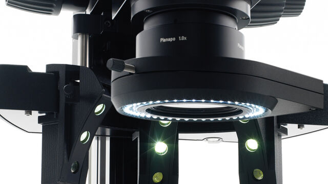
光学显微镜配件
光学显微镜配件
徕卡显微系统提供多种显微镜配件,结合您的需求和预算,量身定制成像系统。欢迎浏览我们的照明系统、目镜、滤镜、滤镜转盘等产品目录。
产品目录 Show subnavigation
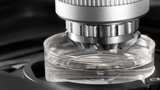
光学显微镜配件
徕卡显微系统提供多种显微镜配件,结合您的需求和预算,量身定制成像系统。欢迎浏览我们的照明系统、目镜、滤镜、滤镜转盘等产品目录。

Leica Science Lab Show subnavigation
阅读我们的最新文章
作为徕卡显微系统有限公司 的信息门户,ScienceLab (徕卡课堂) 提供许多关于显微技术主题的科研和教学材料。其内容宗旨在于为初学者、有经验的从业者和科学家等的日常工作和实验提供支持。

如何通过自动化超薄切片技术节省时间与样本
本文阐述了如何利用树脂包埋电镜样本的 3D micro-CT 数据,在切片前将样本修整至预设目标平面。采用Leica UC Enuity 系统的交互式自动化方案,可显著节省时间、减少样本损耗及缩短新手用户的培训周期。
利用新型可扩展的干细胞培养设计未来
具有远见卓识的生物技术初创企业 Uncommon Bio 正在应对世界上最大的健康挑战之一:食品可持续性。在这次网络研讨会上,干细胞科学家塞缪尔-伊斯特(Samuel East)将展示他们如何使细胞农业的干细胞培养基既安全又经济可行。了解他们如何将培养基成本降低 1000 倍,并开发出不含动物成分、食品安全的 iPSC 培养基。

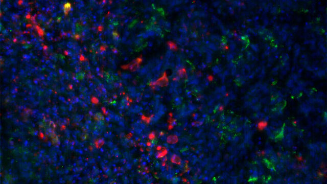
利用大数据探索阿尔茨海默病的空间蛋白组
阿尔茨海默病是一种遗传性和散发性的神经退行性疾病,导致中晚年认知能力下降,特征为β-淀粉样蛋白斑块和 tau蛋白 缠结。由于治疗选择有限,新的研究策略至关重要。Cell DIVE 多重成像解决方案可以对阿尔茨海默病脑组织进行研究,揭示,可能新的研究方向。这里我们展示了 Cell DIVE 多重成像仪的图像查看器,用户能够直接在自己的浏览器中访问完整的阿尔茨海默病多重数据集。

验证汽车零部件的规格
在汽车零部件的开发和生产过程中,无论是供应商还是汽车制造商,都必须符合规格要求。这些规格对保持汽车和其他车辆在生命周期内的性能标准和安全运行至关重要[1,2,3]。在满足或超越日益严格的质量标准的同时,对更高效和更具成本效益的零部件开发和生产的需求一直在提高。本文解释了如何用数码显微镜轻松快速地研究和记录零件以确定其是否符合规格要求。
Mica: 助力伦敦帝国学院开展跨学科科研研究
这篇访谈重点介绍了伦敦帝国学院的 Mica 所产生的变革性影响。科学家们解释了Mica如何改变了游戏规则,扩大了研究的可能性,促进了跨学科合作。他们解释了使用 Mica 进行详细的活细胞成像如何提供更有意义的信息,使科学家始终站在研究的最前沿。研究小组预计,Mica将继续开辟新的研究途径,包括研究微流体技术和其他先进应用。

从显微镜到电镜:完整的冷冻光电联用工作流程
在题为“多模态玻璃化征程,从实验台到电子显微镜的冷冻关联工作流程”的网络研讨会上,专家团队(Edoardo D'Imprima、Zhengyi Yang、Andreia Pinto 和 Martin Fritsch)讨论了光电关联工作流程在结构生物学中的惊艳成果,这些工作流程帮助研究人员研究生物结构的细微细节。接下来让我们一起探索冷冻光电关联(cryo-CLEM)相关工作流程的最新发展、仪器和技术。
癌症研究
癌症是一种复杂的异质性疾病,由于细胞生长失控而引起。 一个或一组细胞的基因和表观遗传的变化破坏了正常功能,导致细胞自发、不受控制地生长和增殖。

如何形成清晰的图像
在显微镜检查中,景深常被看做经验参数。在实际操作中,会根据数值孔径、分辨率和放大率之间的相关性确定该参数。为了获得最佳视觉效果,现代显微镜的调节设备在景深和分辨率之间实现了最佳平衡,这两个参数在理论上呈负相关。

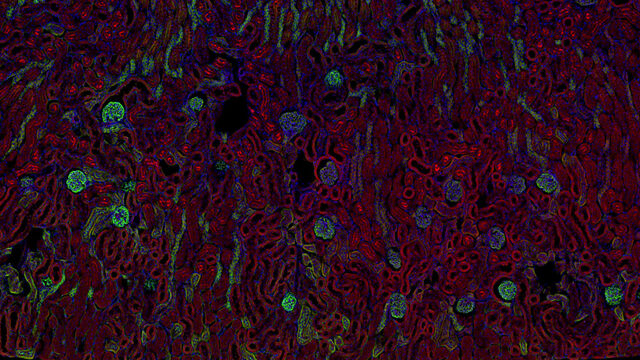
用大数据视角深入了解胰腺癌研究
胰腺癌由于其靠近主要器官难以分辨和难治疗,死亡率接近 40%,。这个研究探讨了胰腺导管腺癌(PDAC)的复杂生物学机制,研究了代谢、凋亡和免疫中肿瘤侵袭性的相关分子结构和空间决定因素。可以访问您的浏览器中的完整 Cell DIVE 数据集,以深入了解这些发现。

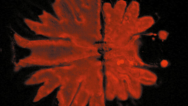
利用大数据查看器揭示结肠癌隐藏的复杂性
结直肠癌是一种的重大健康负担。虽然手术初期有效,但部分患者会发展为预后不良的复发性继发疾病,需要采用免疫疗法等先进治疗手段。利用空间生物学方法,如 Cell DIVE 多重成像技术,可为开发新型治疗方案提供关键洞见。通过 Minerva 图像查看器在浏览器中访问完整的 Cell DIVE 数据集,进一步探索这些发现。
相位对应
使用相差光学显微镜,无需染色就可以更大对比度观察各种类型生物标本的结构。
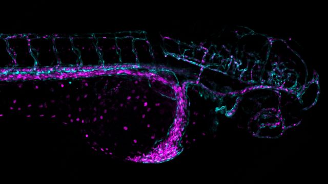
克服显微镜成像移动斑马鱼幼虫时的挑战
Zebrafish is a valuable model organism with many beneficial traits. However, imaging a full organism poses challenges as it is not stationary. Here, this case study shows how zebrafish larvae can be imaged during stationary periods and easily relocated after movement. The seamlessly integrated widefield and confocal capability of Mica is leveraged to capture fast events, like the heartbeat, with virtually no out-of-focus background noise which is inherent to standard widefield systems.
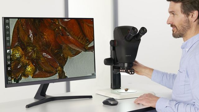
解剖显微镜
实施解剖工作时,您可以通过解剖显微镜的目镜观察很长时间。 徕卡显微系统为您提供各种显微镜和范围广泛的解剖显微镜零配件,确保您能找到最符合您需求的显微镜解决方案。

神经科学研究解决方案
您的工作是更好地了解神经退行性疾病,还是研究神经系统的功能? 了解如何使用徕卡显微系统的成像解决方案取得突破。

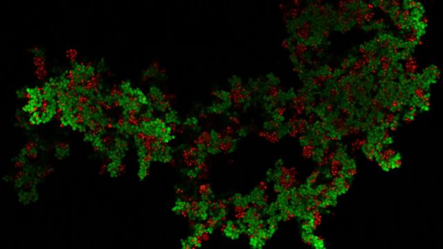
利用人工智能驱动的空间蛋白质组学绘制肿瘤免疫图谱
未经治疗肿瘤的空间图谱分析可呈现肿瘤免疫结构的整体特征,有助于理解治疗反应。具有免疫活性的小鼠模型对于识别肿瘤发生发展过程中免疫依赖性事件至关重要。要表征这些具有完整免疫系统及相互作用细胞组分的模型,需要采用多重标记分析技术。我们展示了一种基于人工智能的空间蛋白质组学方法,用于研究小鼠癌组织中的肿瘤-免疫互作机制。
病毒学
您的主要研究对象是病毒感染和疾病吗? 了解如何使用徕卡显微系统公司的成像和样本制备解决方案深入研究病毒学。


外部滤光片转轮手册
- 高速激发光、衰减、和发射光控制
- 24 ms的切换时间(相邻位置)
- 振动最小
- 设计小巧、紧凑
- 通过强大的软硬件解决方案实现高速同步控制(徕卡AF6000 E、AF6000、AF6500和AF7000)
- 采集速度31 fps
- 可灵活进行不同的设置 - 五位滤盘
- 电动载玻片可插入一体机徕卡EL6000的适配器、显微镜支架或专门的发射光控制C型接口。
- 最多可同时设置四个滤光片。
- 应用包适用于标准荧光、Fura2和FRET。


