
Filter articles
标签
产品
Loading...

Coherent Raman Scattering Microscopy Publication List
CRS (Coherent Raman Scattering) microscopy is an umbrella term for label-free methods that image biological structures by exploiting the characteristic, intrinsic vibrational contrast of their…
Loading...

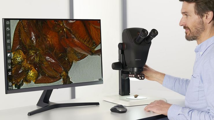
Get to Insights Faster and Easier with AI Image Analysis Tools
Discover how Aivia helps scientists streamline image analysis with fast setup, accurate AI detection, and easy batch processing.
Loading...

Unlocking the Secrets of Organoid Models in Biomedical Research
Get ready to delve deeper into the world of organoids and 3D models, which are essential tools for advancing our understanding of human health. Navigating these complex structures and obtaining clear…

Loading...

临床显微镜:相机选择的考虑因素
过去几年,病理学实验室对图像的需求显著增加,无论是在组织病理学、细胞学、血液学、临床微生物学还是其他应用领域。除了诊断记录外,图像还服务于许多其他目的。然而,通过目镜观察到的图像和数字图像在本质上是不同的,一个是光学图像,另一个是数字图像。从与相机相关的几个方面来审视这一过程,将有助于确保您能够获取所有细节和颜色保真度的图像。
Loading...

选择临床显微镜需考虑的因素
显微镜是病理学家工作流程中不可或缺的一部分。特别是在组织病理学、血液病理学或医学微生物学中,病理学家使用显微镜来高效、可靠地进行诊断。因此,他们经常长时间低头看目镜,可能会因为工作姿势而感到身体不适。
Loading...

如何通过自动化超薄切片技术节省时间与样本
本文阐述了如何利用树脂包埋电镜样本的 3D micro-CT 数据,在切片前将样本修整至预设目标平面。采用Leica UC Enuity 系统的交互式自动化方案,可显著节省时间、减少样本损耗及缩短新手用户的培训周期。
Loading...
 applied. Image courtesy of Samuel East, Uncommon Bio.")
利用新型可扩展的干细胞培养设计未来
具有远见卓识的生物技术初创企业 Uncommon Bio 正在应对世界上最大的健康挑战之一:食品可持续性。在这次网络研讨会上,干细胞科学家塞缪尔-伊斯特(Samuel East)将展示他们如何使细胞农业的干细胞培养基既安全又经济可行。了解他们如何将培养基成本降低 1000 倍,并开发出不含动物成分、食品安全的 iPSC 培养基。
